Freezing the time: Ultrafast Transmission Electron Microscopy
In comparison to other ultrafast methods, UTEM occupies a significant and unique territory for obtaining the types of high spatiotemporal resolution information needed to advances several areas of science. UTEM is especially useful in investigating the dynamics of processes, particularly those far from equilibrium and in extreme environments. This includes identifying intermediate states that occur during complex processes before a system reaches equilibrium enabling us to improve our understanding of fast processes in a variety of scientific topics. The realization of UTEM is a revolutionary step for the in-situ investigation of dynamic processes in materials with high spatiotemporal resolution and the capabilities of UTEM provide an ideal platform to probe dynamic phenomena through a combination of imaging, spectroscopy, and diffraction on their natural length and time scales.

The challenge in applying electron microscopic imaging to dynamic processes is achieving electron pulses of sufficiently high intensity and short duration to freeze the electronic, structural, or chemical motion. The high time resolution of an ultrafast TEM (UTEM) is achieved by creating short electron pulses that are used to illuminate the specimen. A UV laser pulse stimulates photoemission of electrons from a photocathode. The resultant bunch of electrons is then accelerated and sent into the electron-optical column. It is then focused onto the specimen and magnified to produce the image. The response to be studied (chemical reactions in energetic materials, a phase transformation, shock propagation, structural change, chemical reaction, etc.) in the experiment is stimulated by a second laser pulse. This method is called “pump-probe scheme” and the delay between the two laser pulses sets the timing of the observation.
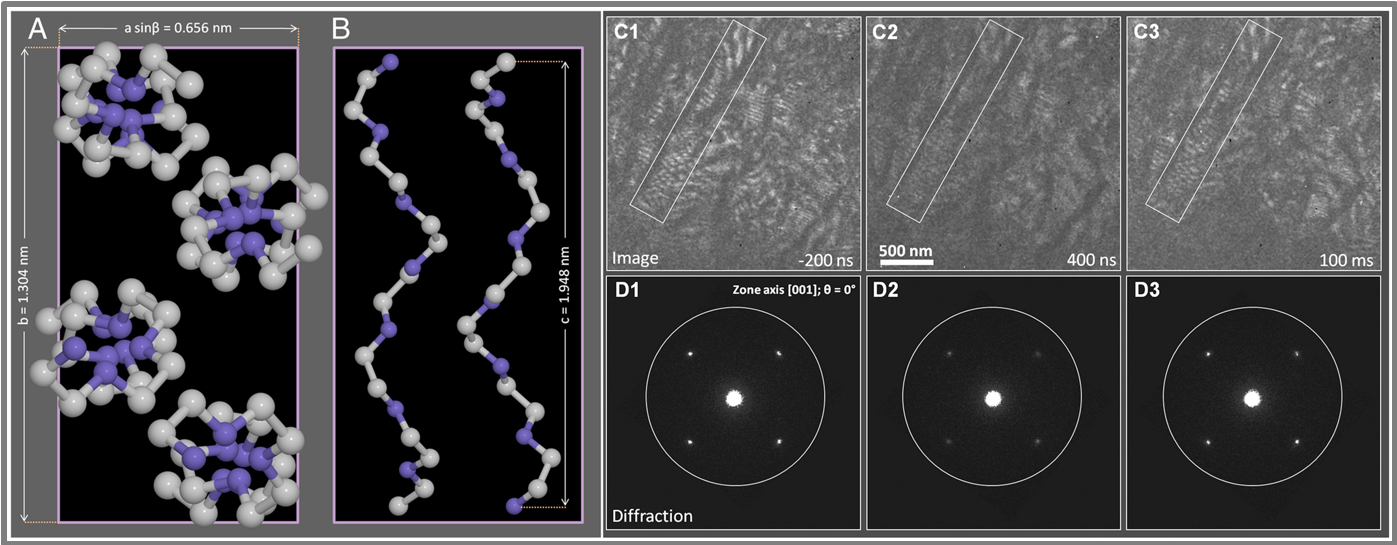
For beam-sensitive specimens, there are stringent requirements on the dose used to form an image or diffraction pattern. Ultrafast transmission electron microscopy has the fastest shutter that enables electron-beam irradiation in a very short exposure time and with accurate control of dosage. Thus, it is possible to achieve imaging on a time scale shorter than that of radiation damage. Therefore, pulsed beam operation of UTEM minimizes the dose and opens completely new avenues to investigate the fundamental properties of soft materials, organic crystals and biological materials. The pulsed imaging mode of the UTEM utilizes doses that are substantially below that for a conventional TEM. For exampe, we have utilized the low dose imaging capabilities of UTEM using pulsed electrons to investigate the structural dynamics of organic polymers. We performed direct imaging of structural dynamics of helical macromolecules of poly(ethylene) oxide (PEO) over the time scales of conformational dynamics (ns to subsecond) by UTEM in the single-shot and stroboscopic modes. With temporally controlled electron dosage, we obtained both diffraction and real-space images without irreversible radiation damage.
We demostrated Ultrafast Scanning Transmission Electron Microscopy (USTEM) in the imaging of silver nano-wires and gold nanoparticles. For the wire, the mechanical motion and shape morphological dynamics were imaged, and from the images we obtained the resonance frequency and the dephasing time of the motion. Moreover, we demonstrate here the simultaneous acquisition of dark-field images and electron energy loss spectra from a single gold nanoparticle, which is not possible with conventional methods. The methodology is wide ranging in applications, particularly in heterogeneous systems, because incoherent imaging (Z2 dependence) makes visualization specific to component elements and to the selected area. The local probing capabilities of USTEM open new avenues for probing dynamic processes, from single isolated to embedded nanostructures, without being affected by the heterogeneous processes of ensemble-averaged dynamics. Such methodology promises to have wide-ranging applications in materials science and in single-particle biological imaging.

